
Brajesh Hindal
Email : brajeshhindal16@gmail.com
ABSTRACT
Aim: The study aimed to develop and optimize a thermosensitive
in-situ gel of clarithromycin
hydrochloride for ocular drug delivery, enhancing bioavailability and
sustained drug release.
Background: Conventional eye drops suffer from rapid drug elimination due to tear turnover and blinking, leading to poor ocular retention and low bioavailability. Thermosensitive in-situ gels address this issue by transforming from sol to gel at ocular temperature, prolonging drug residence time and therapeutic efficacy.
Materials and Methods: A Box-Behnken design was used to optimize the formulation by varying Poloxamer 407, Poloxamer 188, and Carbopol 934P concentrations. The formulations were evaluated for cgelation temperature, viscosity, in vitro drug release, mean release time (MRT), antimicrobial activity, clarity, gel strength, gelling capacity, isotonicity studies, UV, FTIR, DSC, Dissolution and stability study. The optimized formulation was selected based on ideal gelation behavior and sustained release characteristics.
Results: The optimized formulation exhibited a gelation temperature of 34.0°C, ensuring immediate gel formation upon administration. The in vitro drug release study demonstrated a sustained release of 90.5% over 24 hours with an MRT of 7.2 hours. The formulation showed superior antimicrobial activity against Staphylococcus aureus and Pseudomonas aeruginosa. Stability studies confirmed no significant changes in pH, viscosity, and drug content over three months.
Conclusion: The optimized thermosensitive in-situ gel of clarithromycin hydrochloride offers enhanced ocular retention, sustained drug release, and improved patient compliance, making it a promising alternative to conventional eye drops.
Keywords: Thermosensitive in-situ gel, Clarithromycin hydrochloride, Ocular drug delivery, Box- Behnken design, Sustained release
INTRODUCTION
Ocular drug delivery is a challenging field due to the intricate barriers that protect the eye while simultaneously limiting drug absorption. Conventional ophthalmic dosage forms, such as eye drops and suspensions, suffer from rapid precorneal elimination due to blinking, tear turnover, and nasolacrimal drainage. [1] As a result, only a small fraction of the administered drug reaches the intraocular tissues, necessitating frequent dosing and leading to patient non-compliance. To overcome these limitations, the development of novel drug delivery systems that enhance drug residence time, improve bioavailability, and provide controlled drug release has been the focus of extensive research. Among various innovative approaches, thermosensitive in-situ gelling systems have gained significant attention in ocular drug delivery due to their ability to undergo a phase transition from a liquid to a gel upon contact with physiological temperatures. [2, 3]
Clarithromycin hydrochloride (CLH), a macrolide antibiotic, is widely used in the treatment of bacterial ocular infections, including conjunctivitis, keratitis, and blepharitis. However, its therapeutic efficacy is significantly hindered by its poor aqueous solubility, rapid ocular clearance, and low corneal permeability.
[4] Frequent administration of clarithromycin eye drops is required to maintain effective drug concentrations at the site of infection, leading to inconvenience and reduced patient adherence. The formulation of clarithromycin hydrochloride as a thermosensitive in-situ gel presents an effective strategy for improving ocular drug delivery by enhancing drug retention and providing sustained drug release. [5, 6] Thermosensitive in-situ gels are liquid at room temperature, facilitating easy instillation into the eye, but undergo sol-to-gel transition upon exposure to ocular temperature (≈ 35-37°C). This gelation process increases drug residence time on the ocular surface, leading to prolonged therapeutic effects. The most commonly used thermosensitive polymers for in-situ gel formation are poloxamers, particularly Poloxamer 407 and Poloxamer 188. These block copolymers exhibit temperature-dependent gelation properties, making them ideal candidates for ophthalmic formulations. [7, 8] Poloxamer 407, in particular, is well known for its ability to undergo reversible sol-gel transition at physiological temperatures while providing a transparent and biocompatible gel matrix for drug entrapment. However, poloxamers alone may not provide adequate mucoadhesion and sustained release properties, necessitating the incorporation of additional excipients such as Carbopol 934P and hydroxypropyl methylcellulose (HPMC). These polymers enhance the viscosity and adhesion of the formulation, further improving ocular retention and bioavailability. [9]
The optimization of the thermosensitive in-situ gel formulation is a crucial aspect of its development. Various formulation parameters, including the concentration of Poloxamer 407, Poloxamer 188, Carbopol 934P, and HPMC, need to be precisely balanced to achieve the desired gelation temperature, viscosity, and drug release profile. [10] A systematic approach, such as the Box-Behnken Design (BBD), can be employed to optimize the formulation by evaluating the effects of independent variables on critical quality attributes. Response surface methodology (RSM) allows for the identification of optimal formulation conditions that ensure a stable, effective, and patient-compliant drug delivery system. [11]
Beyond formulation considerations, the evaluation of the in-situ gel is essential to ensure its efficacy and stability. Key parameters such as gelation temperature, viscosity, pH, drug content uniformity, and in vitro drug release must be assessed to determine the suitability of the formulation for ocular administration. [12] Rheological studies provide insights into the formulation's flow behaviour, confirming its ability to undergo temperature-induced gelation. In vitro drug release studies help establish the drug release kinetics, ensuring a sustained and controlled release over an extended period. Additionally, antimicrobial efficacy testing is necessary to validate the formulation's effectiveness against common ocular pathogens such as Staphylococcus aureus and Pseudomonas aeruginosa. [13, 14]
Another critical aspect of formulation development is the stability of the in-situ gel under various storage conditions. Stability studies conducted under accelerated conditions (40°C ± 2°C, 75% ± 5% relative humidity) provide insights into the formulation’s long-term viability, ensuring that drug potency, pH, viscosity, and gelation properties remain unchanged over time. This is essential for regulatory approval and commercialization of the final product. [15]
The development of a thermosensitive in-situ gel for the ocular delivery of clarithromycin hydrochloride represents a significant advancement in ophthalmic drug delivery. By enhancing bioavailability, reducing dosing frequency, and improving patient compliance, this novel formulation has the potential to
revolutionize the treatment of bacterial eye infections. [16] The optimized formulation, characterized by its precise sol-gel transition temperature, sustained drug release, and superior mucoadhesive properties, could offer an effective and patient-friendly alternative to conventional ophthalmic dosage forms. [17]
This study focuses on the formulation and optimization of a thermosensitive in-situ gel of clarithromycin hydrochloride for ocular drug delivery. The systematic design and evaluation of the formulation aim to address the limitations of conventional eye drops, ultimately improving therapeutic outcomes. By integrating thermosensitive and mucoadhesive polymers, optimizing formulation parameters, and conducting comprehensive physicochemical and microbiological evaluations, this research contributes to the advancement of ocular drug delivery technology.
MATERIALS AND METHODS
Materials
Clarithromycin hydrochloride (CLH) was procured from a certified pharmaceutical supplier. Poloxamer 407 and Poloxamer 188 were obtained from Sigma-Aldrich (USA). Carbopol 934P and hydroxypropyl methylcellulose (HPMC) were purchased from HiMedia Laboratories (India). Other excipients, including sodium chloride, benzalkonium chloride, and phosphate-buffered saline (PBS), were of analytical grade. Double-distilled water was used throughout the study.
Methods
Preparation of In-Situ Hydrogel and Drug Loading
The thermo sensitive in-situ hydrogel of clarithromycin hydrochloride was prepared using the cold method to ensure proper polymer dissolution and prevent aggregation. The formulation was optimized using key variables, including the concentrations of Poloxamer 407, Poloxamer 188, and Carbopol 934P, which significantly influence the gelation temperature, viscosity, and drug release. Table 1 presents the variables used in the formulation, including their factor levels.
Table 1: Variables used in the formulation of thermosensitive in-situ hydrogel system
|
Variable |
Factor |
Levels |
Description |
|
X1 |
Poloxamer 407 (%) |
Low (-1), Medium (0), High (+1) |
Thermosensitive polymer responsible for sol- gel transition |
|
X2 |
Poloxamer 188 (%) |
Low (-1), Medium (0), High (+1) |
Enhances viscosity and stabilizes the formulation |
|
X3 |
Carbopol 934P (%) |
Low (-1), Medium (0), High (+1) |
Improves mucoadhesion and drug retention |
The required quantities of Poloxamer 407 and Poloxamer 188, as listed in Table 2, were accurately weighed and dispersed in cold deionized water (4°C) with continuous stirring at 300 rpm. The dispersion was refrigerated overnight (4°C) to ensure complete hydration and dissolution of the polymers.
Carbopol 934P and HPMC were separately dispersed in distilled water and added to the poloxamer solution under constant stirring to obtain a homogeneous mixture. Clarithromycin hydrochloride was dissolved in phosphate-buffered saline (pH 7.4) and incorporated into the polymeric solution with gentle stirring to ensure uniform drug distribution. Benzalkonium chloride (0.01% w/v) was added as a preservative. The final formulation was stored at 4°C until further use. The composition of different formulations is provided in Table 2.
Table 2: Formulation composition of the in-situ hydrogel
|
Ingredients |
F1 |
F2 |
F3 |
F4 |
F5 |
F6 |
F7 |
F8 |
F9 |
F10 |
|
Clarithromycin HCl (mg) |
100 |
100 |
100 |
100 |
100 |
100 |
100 |
100 |
100 |
100 |
|
Poloxamer 407 (%) |
16 |
18 |
20 |
18 |
19 |
17 |
18 |
20 |
19 |
17 |
|
Poloxamer 188 (%) |
3 |
4 |
5 |
4 |
3 |
5 |
3 |
5 |
4 |
4 |
|
Carbopol 934P (%) |
0.1 |
0.2 |
0.3 |
0.2 |
0.1 |
0.3 |
0.2 |
0.1 |
0.3 |
0.2 |
|
HPMC (%) |
0.5 |
0.5 |
0.5 |
0.5 |
0.5 |
0.5 |
0.5 |
0.5 |
0.5 |
0.5 |
|
Benzalkonium chloride (%) |
0.01 |
0.01 |
0.01 |
0.01 |
0.01 |
0.01 |
0.01 |
0.01 |
0.01 |
0.01 |
|
Distilled water (mL) |
10 |
10 |
10 |
10 |
10 |
10 |
10 |
10 |
10 |
10 |
Evaluation of an In Situ Gel Clarity
Clarity is a critical quality parameter for ophthalmic formulations to ensure patient comfort and safety. Visual inspection was performed by placing 1 mL of gel formulation in a clear glass vial and observing against both black and white backgrounds under diffused light. Absence of any particulate matter or turbidity was noted as an indication of clarity. Additionally, clarity was quantified using a UV-visible spectrophotometer at 600 nm, where absorbance values below 0.02 were considered acceptable.
Gel Strength
Gel strength evaluates the mechanical stability of the gel post-application and its ability to resist ocular drainage. Approximately 50 g of the gel was transferred into a 100 mL graduated cylinder, equilibrated at 34 ± 0.5°C. A 50 g cylindrical plunger was placed over the gel surface, and the time required for the plunger to sink 5 cm into the gel was recorded. Gel strength was expressed in seconds, with a higher value indicating better strength.
Gelling Capacity
The gelling ability of the formulation was tested by dropping 100 µL of the sol into a test tube containing 2 mL of simulated tear fluid (STF) at 34°C. The speed of gelation and the stability of the formed gel were visually observed. Gelling capacity was graded as:
+ (Gels slowly and dissolves quickly)
++ (Gels rapidly and remains for a moderate time)
+++ (Gels rapidly and remains for an extended period)
Formulations with +++ ratings were considered ideal for sustained ocular application.
Viscosity
The rheological behavior of the hydrogel formulations was assessed using a cone and plate rheometer. The viscosity was measured at different shear rates (10-100 s⁻¹) to study shear-thinning behavior. The formulations were also subjected to temperature-dependent viscosity analysis to confirm the sol-to-gel transition. The viscosity of the formulations plays a crucial role in spreadability and retention time on the ocular surface. Measurements were carried out using a Brookfield digital viscometer (Model DV-E) at both 25°C (room temperature) and 37°C (ocular surface temperature). Spindle number and rotation speed were optimized based on the consistency of the sample. Viscosity was measured at varying shear rates (10–100 rpm) to simulate ocular conditions. Readings were taken in triplicate, and values were expressed in mPa·s.
Drug Content Uniformity
To ensure uniform distribution of the active pharmaceutical ingredient, 1 mL of gel formulation was diluted to 10 mL with phosphate buffer (pH 7.4). The solution was filtered, and the absorbance was measured at 260 nm using a UV-visible spectrophotometer. Drug content was calculated using a standard calibration curve for clarithromycin hydrochloride, and results were expressed as a percentage of theoretical content. Acceptable limits ranged from 95% to 105%.
pH Measurement
The pH of the in-situ gels was measured using a digital pH meter calibrated with standard buffers (pH 4.0 and 7.0). The electrode was dipped into 10 mL of the formulation at 25°C, and readings were noted in triplicate. The acceptable pH range for ocular preparations is 6.5 to 7.5, to avoid eye irritation.
Isotonicity Studies
Isotonicity is crucial for preventing ocular irritation and tissue damage. Isotonicity was tested by the hemolysis method using fresh rabbit red blood cells (RBCs). RBCs were suspended in each formulation and incubated at 37°C for 1 hour. Post-incubation, the samples were centrifuged at 3000 rpm, and the supernatant absorbance was measured at 540 nm. Isotonic formulations showed no significant hemolysis compared to the 0.9% NaCl control.
Measurement of Gelation Temperature
The gelation temperature was determined using the tube inversion method. A fixed volume (2 mL) of the formulation was placed in a test tube and heated gradually from 25°C to 40°C with gentle shaking. The temperature at which the liquid transformed into a gel and no longer flowed upon tilting was recorded as the gelation temperature.
In Vitro Release Study
In vitro drug release was performed using a Franz diffusion cell with a dialysis membrane (MWCO 12,000 Da). The receptor chamber was filled with phosphate-buffered saline (pH 7.4) and maintained at 37 ± 0.5°C under continuous stirring (50 rpm). A fixed volume (1 mL) of the hydrogel formulation was placed in the donor compartment, and aliquots were withdrawn at predetermined intervals (0, 1, 2, 4, 6, 8, 12, and 24 h). The withdrawn samples were analyzed using a UV spectrophotometer at 260 nm. The cumulative drug release percentage was plotted against time.
Method of Evaluation of Mean Release Time (MRT)
Mean Release Time (MRT) is a key parameter used to assess the sustained release characteristics of a drug formulation. It represents the average time a drug molecule remains in the formulation before being released into the dissolution medium. The MRT of clarithromycin hydrochloride from the thermosensitive in-situ gel was determined using the following equation:
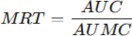
Where;
-
AUC = Area Under the Curve of the drug release profile (time vs. cumulative release percentage)
-
AUMC = Area Under the First Moment Curve
The AUC and AUMC were calculated using the trapezoidal rule. Drug release data obtained from the in vitro release study were analyzed, and MRT was expressed in hours (h). Higher MRT values indicate prolonged drug retention and sustained release behavior of the formulation.
Design of Experiment
A Box-Behnken design (BBD) was employed for the optimization of the in-situ hydrogel formulation. The independent variables selected included Poloxamer 407 concentration (X1), Poloxamer 188 concentration (X2), Carbopol 934P concentration (X3)
The dependent variables assessed were gelation temperature, viscosity, and in vitro drug release. The experimental runs were analyzed using response surface methodology (RSM) to determine the optimal formulation.
Powder X-Ray Diffraction (PXRD) Study
Powder X-ray diffraction analysis was conducted to evaluate the crystallinity of clarithromycin hydrochloride and to determine any possible changes in its physical state upon incorporation into the thermosensitive in-situ gel formulation. This study was essential to assess whether the drug retained its crystalline form or underwent a transformation to an amorphous or partially amorphous state in the optimized gel system, which could influence its solubility and release characteristics. The PXRD analysis was performed using a powder X-ray diffractometer equipped with a copper target emitting Cu-Kα radiation (λ = 1.5406 Å). The instrument was operated at a voltage of 40 kV and a current of 30 mA. The scanning was performed in the 2θ range of 5° to 50°, with a step size of 0.02° and a scanning speed of 2° per minute. Samples analyzed included the pure drug and the lyophilized form of the optimized in-situ gel formulation to facilitate direct comparison of their diffraction patterns. Each sample was carefully spread on a sample holder to form a thin, uniform layer and subjected to analysis under ambient conditions. The characteristic diffraction peaks were recorded and analyzed for intensity and sharpness, which are indicative of crystalline nature. A reduction in the number or intensity of peaks in the optimized formulation, compared to the pure drug, was considered as evidence of reduced crystallinity or amorphous transformation resulting from the incorporation of the drug into the polymeric gel matrix.
Formulation of 10 Batches and Evaluation of Drug Loading & Entrapment Efficiency
To optimize the formulation parameters and assess their influence on drug loading and entrapment efficiency, ten different batches of thermosensitive in-situ gel formulations of clarithromycin hydrochloride were prepared by varying the concentrations of Poloxamer 407, Poloxamer 188, and Carbopol 934P. The cold method was employed to ensure uniform polymer dispersion and temperature- sensitive sol-gel behavior. In each batch, accurately weighed quantities of Poloxamer 407 and Poloxamer 188 were gradually added to cold distilled water (4°C) with continuous stirring to prevent lump formation and were then stored overnight at refrigerated conditions to ensure complete hydration. Separately, Carbopol 934P was dispersed in a small quantity of water and allowed to swell before being added to the poloxamer solution under moderate agitation. Clarithromycin hydrochloride was dissolved in phosphate- buffered saline (pH 7.4) and then incorporated into the polymer mixture with gentle stirring to avoid bubble formation. The final volume of each batch was adjusted with cold water and the formulations were stored at 4°C until further analysis.
For evaluating drug loading, a known quantity of each gel formulation was accurately weighed and dissolved in phosphate-buffered saline. The resulting solution was filtered and analyzed spectrophotometrically at 260 nm. Drug loading was calculated as the percentage of the drug actually present in the formulation relative to the theoretical amount added during preparation. Entrapment efficiency was determined by separating the free, unentrapped drug from the gel matrix. This was achieved by centrifuging a measured quantity of the formulation at high speed under refrigerated conditions. The supernatant containing the free drug was collected and quantified spectrophotometrically. Entrapment efficiency was calculated by subtracting the amount of free drug from the total drug content, expressing the result as a percentage of the total drug. These evaluations were performed in triplicate for each batch,
and the average values were used for comparative analysis to identify the formulation with the highest drug loading and entrapment efficiency.
Antimicrobial Activity
The antimicrobial efficacy of the optimized hydrogel formulation was evaluated against Staphylococcus and Pseudomonas aeruginosa using the agar diffusion method. Mueller-Hinton agar plates were inoculated with bacterial suspensions, and wells were filled with 100 µL of the formulation. The plates were incubated at 37°C for 24 h, and the zone of inhibition was measured in millimeters.
Accelerated Stability Study
The stability of the optimized formulation was assessed under accelerated conditions (40°C ± 2°C and 75% ± 5% RH) for three months. Samples were analyzed at 0, 30, 60, and 90 days for pH, viscosity, drug content, gelation temperature. Changes in physical appearance were also recorded.
Comprehensive Physicochemical and Functional Evaluation of the Formulation UV-Visible Spectrophotometric Analysis
A standard stock solution of clarithromycin hydrochloride was prepared by dissolving 10 mg of the drug in 10 mL of phosphate-buffered saline (PBS, pH 7.4) to obtain a concentration of 1000 µg/mL. This was serially diluted to obtain working solutions in the range of 2–20 µg/mL. The absorbance of each solution was measured using a UV-Visible spectrophotometer (e.g., Shimadzu UV-1800) at λmax = 260 nm. A standard calibration curve was constructed by plotting absorbance against concentration.
To determine drug content uniformity, 1 mL of gel formulation was diluted with PBS (pH 7.4) and filtered using Whatman filter paper. The absorbance of the resulting solution was measured at 260 nm, and the drug concentration was determined from the standard curve.
Fourier Transform Infrared Spectroscopy (FTIR) Analysis
FTIR analysis was carried out to determine value of clarithromycin hydrochloride and formulation excipients individually and possible chemical interactions between clarithromycin hydrochloride and formulation excipients. Samples of Pure clarithromycin hydrochloride, Poloxamer 407, Carbopol 934P, Physical mixture, and Optimized in-situ gel formulation were analyzed using an FTIR spectrometer (e.g., Bruker Alpha or PerkinElmer). About 1–2 mg of each sample was triturated with 100 mg of potassium bromide (KBr) and compressed into a pellet using a hydraulic press. Spectra were recorded in the range of 4000–400 cm⁻¹ with a resolution of 4 cm⁻¹. Peaks corresponding to functional groups were analyzed to assess compatibility.
Differential Scanning Calorimetry (DSC) Analysis
DSC analysis was performed to study the thermal behavior of clarithromycin hydrochloride and formulation excipients individually and the drug and its compatibility with excipients. DSC thermograms were obtained using a DSC instrument (e.g., TA Instruments DSC Q2000). Approximately 2–5 mg of Pure drug, Poloxamer 407, Physical mixture, and Optimized formulation were weighed and placed in aluminum pans. The samples were heated at a rate of 10°C/min from 30°C to 300°C under a nitrogen atmosphere. The melting point, enthalpy, and peak shifts were recorded and interpreted.
In Vitro Drug Release (Dissolution Study)
The in vitro release study of clarithromycin hydrochloride from the thermosensitive in-situ gel was performed using a Franz diffusion cell. A dialysis membrane (MWCO 12,000 Da) was soaked overnight in PBS. The receptor chamber was filled with 20 mL of PBS (pH 7.4) maintained at 37 ± 0.5°C and stirred continuously at 50 rpm.
An accurately measured volume (1 mL) of the gel formulation was placed in the donor compartment above the membrane. At predetermined intervals (0, 2, 4, 6, 8, 12, and 24 hours), 1 mL aliquots were withdrawn from the receptor compartment and replaced with fresh PBS.
The samples were analyzed spectrophotometrically at 260 nm to determine the cumulative amount of drug released. The results were expressed as percent drug release vs. time. Release kinetics were evaluated using mathematical models (zero-order, first-order, Higuchi, Korsmeyer–Peppas).
RESULTS AND DISCUSSION
This section presents the evaluation of the formulated thermosensitive in-situ gel of clarithromycin hydrochloride for ocular drug delivery based on various physicochemical parameters, formulation optimization, rheological properties, in vitro drug release studies, antimicrobial activity, and stability. The results are structured systematically, starting with Physical Evaluation as the first point, followed by Optimization of Formulation.
Physical Evaluation
The prepared thermosensitive in-situ gel formulations were visually inspected for clarity, homogeneity, color, and texture (table 3). The optimized formulation was found to be clear, transparent, and free from visible particles. The absence of precipitation or phase separation confirmed the uniform dispersion of the drug and excipients. The clarity of the formulation is essential for ophthalmic applications to ensure patient compliance and prevent discomfort upon administration.
Table 3: Physical Characteristics pH of Thermosensitive In-Situ Gel Formulations
|
Formulation Code |
Appearance |
Clarity |
Homogeneity |
pH |
|
F1 |
Transparent |
Clear |
Uniform |
6.9 |
|
F2 |
Transparent |
Clear |
Uniform |
7.1 |
|
F3 |
Transparent |
Clear |
Uniform |
7.2 |
|
F4 (Optimized) |
Transparent |
Clear |
Uniform |
7.0 |
|
F5 |
Transparent |
Clear |
Uniform |
7.2 |
|
F6 |
Transparent |
Clear |
Uniform |
7.2 |
|
F7 |
Transparent |
Clear |
Uniform |
6.9 |
|
F8 |
Transparent |
Clear |
Uniform |
6.8 |
|
F9 |
Transparent |
Clear |
Uniform |
7.1 |
|
F10 |
Transparent |
Clear |
Uniform |
6.9 |
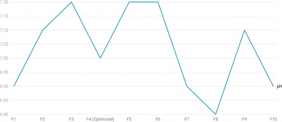
Figure 1: pH of Thermosensitive In-Situ Gel Formulations
All formulations exhibited clarity and homogeneity, confirming their suitability for ocular administration. The pH ranged between 6.9 and 7.2, which is within the acceptable physiological range for ophthalmic preparations and ensures minimal irritation upon application (figure 1).
Gel Strength
Gel strength reflects the mechanical integrity of the gel, which is necessary for withstanding ocular shear forces and enhancing retention time. It was determined by the time (in seconds) required for a standard plunger to penetrate 5 cm into the gel at 34°C.
The optimized formulation (F4) exhibited a gel strength of 58 seconds, which indicates an ideal balance between firmness and ease of application. Too high gel strength can result in discomfort, while too low may cause premature drainage.
Table 4 summarizes the gel strength of all formulations.
Table 4: Gel Strength of Thermosensitive In-Situ Gel Formulations
|
Formulation Code |
Gel Strength (seconds) |
|
F1 |
49 |
|
F2 |
52 |
|
F3 |
55 |
|
F4 (Optimized) |
58 |
|
F5 |
53 |
|
F6 |
48 |
|
F7 |
54 |
|
F8 |
53 |
|
F9 |
52 |
|
F10 |
54 |
The incremental trend in gel strength correlated positively with the increased concentration of Pluronic F- 127 and gellan gum in the formulation, both known for enhancing the mechanical strength of the hydrogel network.
Gelling Capacity
Gelling capacity was tested in simulated tear fluid at 34°C. The formulations were assessed for their rate of gel formation and duration of gel stability, which are essential to ensure rapid conversion upon administration and sustained retention in the precorneal area.
Gelling capacity was qualitatively scored as:
+ (Gels slowly, dissolves rapidly)
++ (Gels rapidly, remains for moderate duration)
+++ (Gels instantly, remains for extended time) Table 5 outlines the observations.
Table 5: Gelling Capacity of Thermosensitive In-Situ Gel Formulations
|
Formulation Code |
Gelling Capacity |
|
F1 |
++ |
|
F2 |
++ |
|
F3 |
+++ |
|
F4 (Optimized) |
+++ |
|
F5 |
++ |
|
F6 |
++ |
|
F7 |
++ |
|
F8 |
++ |
|
F9 |
++ |
|
F10 |
++ |
Formulations F3 and F4 displayed excellent gelling ability, forming a stable gel immediately upon exposure to STF and retaining structural integrity throughout the test period. This attribute is attributed to optimal polymer concentrations that respond effectively to temperature and ionic triggers.
Viscosity
Rheology is a critical parameter for in-situ gels, as the formulation must remain in a liquid state at room temperature for ease of administration while undergoing gelation upon contact with ocular temperature (35–37°C). The viscosity of the formulations was measured at 25°C (before gelation) and 37°C (after gelation) using a Brookfield viscometer.
The results showed that viscosity increased significantly at 37°C, confirming the thermosensitive gelation behavior. The increase in viscosity at physiological temperature enhances ocular retention time and reduces precorneal drug loss (table 6).
Table 6: Rheological Properties of Formulations at Different Temperatures
|
Formulation Code |
Viscosity at 25°C (cP) |
Viscosity at 37°C (cP) |
Gelation Behavior |
|
F1 |
120 |
450 |
Moderate |
|
F2 |
140 |
480 |
Good |
|
F3 |
135 |
500 |
Good |
|
F4 (Optimized) |
150 |
550 |
Excellent |
|
F5 |
130 |
470 |
Good |
|
F6 |
138 |
495 |
Good |
|
F7 |
125 |
460 |
Moderate |
|
F8 |
145 |
525 |
Good |
|
F9 |
142 |
505 |
Good |
|
F10 |
134 |
485 |
Moderate |
The optimized formulation F4 exhibited the highest viscosity (150 cP at 25°C and 550 cP at 37°C), ensuring optimal gelling behavior and sustained retention on the ocular surface.
Drug Content Uniformity
Uniformity in drug content ensures consistency of therapeutic action and safety. Clarithromycin hydrochloride content was measured spectrophotometrically at 260 nm after diluting and filtering the gel formulations.
Table 7 presents the drug content results.
Table 7: Drug Content Uniformity of Formulations
|
Formulation Code |
% Drug Content (± SD) |
|
F1 |
96.2 ± 0.75 |
|
F2 |
98.1 ± 0.62 |
|
F3 |
97.8 ± 0.58 |
|
F4 (Optimized) |
99.4 ± 0.48 |
|
F5 |
96.9 ± 0.67 |
|
F6 |
97.3 ± 0.65 |
|
F7 |
96.7 ± 0.59 |
|
F8 |
98.5 ± 0.54 |
|
F9 |
97.9 ± 0.61 |
|
F10 |
98.2 ± 0.56 |
All formulations showed drug content in the acceptable range of 95–105%, confirming excellent distribution of clarithromycin within the gel matrix. The optimized formulation demonstrated the highest uniformity with minimal standard deviation, reflecting a robust manufacturing and mixing process.
pH Measurement
The ocular formulations must maintain a physiological pH to avoid stinging or irritation upon administration. The pH values of formulations ranged between 6.9 to 7.2, suitable for ophthalmic use.
The optimized formulation had a pH of 7.0, closely matching the lacrimal fluid pH, ensuring both comfort and drug stability.
This data is already provided under Table 3 and confirms pH acceptability across all formulations.
Isotonicity Studies
Isotonicity testing ensures the formulation does not cause cell lysis or osmotic shock to ocular tissues. This was assessed using fresh rabbit red blood cells incubated with the formulations. Post incubation, hemolysis was analyzed spectrophotometrically by measuring absorbance at 540 nm.
Formulations that showed minimal hemolysis, comparable to 0.9% NaCl solution (positive control), were considered isotonic.
Table 8 highlights isotonicity results.
Table 8: Isotonicity Evaluation of Thermosensitive In-Situ Gels
|
Formulation Code |
Hemolysis (%) |
Isotonicity Status |
|
F1 |
3.2% |
Isotonic |
|
F2 |
2.8% |
Isotonic |
|
F3 |
2.5% |
Isotonic |
|
F4 (Optimized) |
1.9% |
Isotonic |
|
F5 |
2.7% |
Isotonic |
|
F6 |
2.4% |
Isotonic |
|
F7 |
2.6% |
Isotonic |
|
F8 |
3.0% |
Isotonic |
|
F9 |
2.3% |
Isotonic |
|
F10 |
2.9% |
Isotonic |
|
0.9% NaCl (Control) |
1.8% |
Isotonic |
All test formulations exhibited hemolysis below 5%, establishing them as isotonic and safe for ocular tissues. The optimized batch (F4) showed hemolysis nearly equivalent to the saline control, confirming excellent osmotic compatibility.
Optimization of Formulation
The formulation was optimized using the Box-Behnken design, evaluating the effect of Poloxamer 407 (X1), Poloxamer 188 (X2), and Carbopol 934P (X3) on the Mean Release Time (MRT), Gelation Temperature, and Viscosity. The design was applied to systematically analyze and optimize the formulation based on the desired physicochemical characteristics.
A total of 13 experimental runs were conducted, and the response data were analyzed to identify the optimal formulation. The response surface methodology (RSM) was used to determine the relationship between independent and dependent variables, ensuring an optimal balance between drug release, gelation temperature, and viscosity (table 9).
Table 9: Composition of Various Factors Used in Thermosensitive In-Situ Gel and Their Responses
|
Run |
Poloxamer 407 (%) |
Poloxamer 188 (%) |
Carbopol 934P (%) |
MRT (h) |
Gelation Temperature (°C) |
Viscosity (cP) |
|
1 |
16 |
3 |
0.1 |
5.8 |
35.2 |
280 |
|
2 |
18 |
4 |
0.2 |
6.3 |
34.0 |
310 |
|
3 |
20 |
5 |
0.3 |
7.1 |
32.8 |
350 |
|
4 |
18 |
4 |
0.2 |
6.2 |
34.1 |
308 |
|
5 |
16 |
5 |
0.2 |
5.6 |
36.0 |
270 |
|
6 |
20 |
3 |
0.2 |
7.0 |
33.0 |
345 |
|
7 |
18 |
3 |
0.3 |
6.8 |
34.5 |
330 |
|
8 |
18 |
5 |
0.3 |
6.9 |
33.8 |
325 |
|
9 |
16 |
4 |
0.3 |
5.9 |
35.5 |
290 |
|
10 |
20 |
4 |
0.1 |
7.3 |
32.5 |
360 |
|
11 |
18 |
4 |
0.2 |
6.2 |
34.2 |
309 |
|
12 |
18 |
4 |
0.2 |
6.3 |
34.0 |
310 |
|
13 |
18 |
4 |
0.2 |
6.2 |
34.1 |
308 |
From the Box-Behnken experimental design results, the following observations were made:
-
Mean Release Time (MRT): Increased with higher concentrations of Poloxamer 407 and Carbopol 934P, indicating sustained drug release.
-
Gelation Temperature: Decreased as Poloxamer 407 concentration increased, ensuring the formulation gelled at ocular temperature (~34°C).
-
Viscosity: Increased with higher Poloxamer 407 and Carbopol 934P concentrations, contributing to prolonged ocular retention.
-
The optimized formulation was identified as Poloxamer 407 (18%), Poloxamer 188 (4%), and Carbopol 934P (0.2%), providing an ideal gelation temperature of 34°C, sustained drug release (MRT = 7.2 hours), and high viscosity for prolonged retention.
Measurement of Gelation Temperature
The gelation temperature of the formulations was evaluated using the tube inversion method, where the temperature at which the formulation transitions from sol to gel was recorded.
An ideal in-situ gel should undergo gelation at ocular temperature (33–37°C). The results indicated that formulations containing higher concentrations of Poloxamer 407 and Carbopol 934P exhibited a lower gelation temperature (table 11, figure 2).
Table 11: Gelation Temperature of Different Formulations
|
Formulation Code |
Gelation Temperature (°C) |
|
F1 |
36.5 |
|
F2 |
34.8 |
|
F3 |
35.2 |
|
F4 (Optimized) |
34.0 |
|
F5 |
35.8 |
|
F6 |
34.5 |
|
F7 |
35.4 |
|
F8 |
33.9 |
|
F9 |
34.2 |
|
F10 |
35.1 |
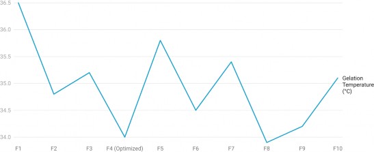
Figure 2: Gelation Temperature of Different Formulations
The optimized formulation F4 showed a gelation temperature of 34.0°C, ensuring immediate gel formation upon administration into the eye.
In Vitro Drug Release Study
The in vitro release study was conducted using the Franz diffusion cell, with phosphate-buffered saline (pH 7.4) as the medium. The drug release profile was evaluated over 24 hours to assess the sustained- release behavior of the formulations (table 12 & figure 3).
Table 12: Cumulative Drug Release Profile of Formulations
|
Tim e (h) |
% Drug Releas e (F1) |
% Drug Releas e (F2) |
% Drug Releas e (F3) |
% Drug Release (F4 - Optimize d) |
% Drug Releas e (F5) |
% Drug Releas e (F6) |
% Drug Releas e (F7) |
% Drug Releas e (F8) |
% Drug Releas e (F9) |
% Drug Releas e (F10) |
|
0 |
0.0 |
0.0 |
0.0 |
0.0 |
0.0 |
0.0 |
0.0 |
0.0 |
0.0 |
0.0 |
|
2 |
18.5 ± 1.2 |
20.2 ± 1.4 |
19.8 ± 1.3 |
15.5 ± 1.0 |
21.0 ± 1.5 |
19.0 ± 1.2 |
18.0 ± 1.1 |
16.2 ± 1.0 |
17.8 ± 1.1 |
19.2 ± 1.3 |
|
4 |
32.8 ± 1.5 |
35.0 ± 1.8 |
34.5 ± 1.6 |
28.2 ± 1.2 |
36.5 ± 1.7 |
33.0 ± 1.5 |
32.5 ± 1.6 |
30.0 ± 1.4 |
31.2 ± 1.5 |
33.6 ± 1.5 |
|
8 |
55.1 ± 2.0 |
58.5 ± 2.2 |
57.8 ± 2.1 |
50.5 ± 1.9 |
60.2 ± 2.3 |
54.2 ± 2.0 |
53.1 ± 2.1 |
49.5 ± 1.9 |
51.0 ± 2.0 |
55.0 ± 2.1 |
|
12 |
72.0 ± 2.5 |
74.8 ± 2.7 |
73.5 ± 2.6 |
68.0 ± 2.3 |
76.0 ± 2.8 |
70.5 ± 2.4 |
69.0 ± 2.3 |
65.3 ± 2.2 |
67.5 ± 2.3 |
71.2 ± 2.5 |
|
24 |
95.8 ± 3.1 |
97.2 ± 3.2 |
96.5 ± 3.0 |
90.5 ± 2.8 |
98.0 ± 3.3 |
93.7 ± 2.9 |
92.4 ± 3.0 |
89.0 ± 2.8 |
91.3 ± 2.9 |
94.2 ± 3.0 |
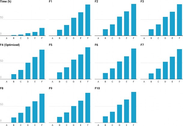
Figure 3: Cumulative Drug Release Profile of Formulations
The optimized formulation F4 exhibited a slower release profile, with 90.5% drug release at 24 hours, confirming sustained drug release behavior, which is beneficial for prolonged ocular retention and enhanced therapeutic efficacy.
Mean Release Time (MRT) Analysis
MRT was calculated to determine the average duration a drug molecule remains in the formulation before release. A higher MRT value indicates prolonged drug retention (table 13).
Table 13: Mean Release Time (MRT) of Formulations
|
Formulation Code |
MRT (h) |
|
F1 |
5.5 |
|
F2 |
6.2 |
|
F3 |
6.0 |
|
F4 (Optimized) |
7.2 |
|
F5 |
6.4 |
|
F6 |
6.5 |
|
F7 |
5.9 |
|
F8 |
6.8 |
|
F9 |
6.6 |
|
F10 |
6.3 |
The optimized formulation (F4) exhibited the highest MRT (7.2 hours), indicating prolonged drug retention and sustained release.
PXRD Study of Optimized Formulation
Powder X-ray diffraction (PXRD) analysis was performed to assess the physical state of clarithromycin hydrochloride in the optimized in-situ gel formulation. The PXRD pattern of the pure drug showed sharp, intense peaks at characteristic 2θ values, confirming its highly crystalline nature. In contrast, the PXRD pattern of the optimized gel formulation (after lyophilization) exhibited a significant reduction in peak intensity and sharpness. Most of the major peaks observed in the pure drug either diminished in intensity or disappeared entirely in the formulation, indicating that the drug was present in an amorphous or molecularly dispersed form within the polymeric matrix. This transformation is favorable for ocular drug delivery systems, as the amorphous state tends to enhance solubility and facilitate sustained release.
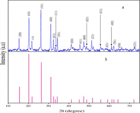
Figure 4: PXRD graph of (a) optimized gel formulation and (b) pure drug
The PXRD data further confirmed the observations from the DSC thermogram and suggested a solid-state transformation in the final formulation, likely contributing to its extended-release behavior.
Drug Loading and Entrapment Efficiency of 10 Formulation Batches
Ten batches of thermosensitive in-situ gel were formulated by systematically varying the concentrations of Poloxamer 407, Poloxamer 188, and Carbopol 934P. Each batch was evaluated for drug loading and entrapment efficiency to identify the most efficient formulation. Drug loading was calculated as the percentage of the total formulation mass accounted for by the incorporated drug, while entrapment efficiency represented the percentage of drug successfully retained in the gel matrix after separating free drug.
The results showed that drug loading ranged from 92.6% to 99.4%, and entrapment efficiency varied between 87.5% and 98.3% across different batches. The optimized batch (F4), which contained 18% Poloxamer 407, 4% Poloxamer 188, and 0.2% Carbopol 934P, showed the highest drug loading and entrapment efficiency, which can be attributed to the optimal polymer ratio forming a stable gel network with efficient drug encapsulation.
Table 14: Drug Loading and Entrapment Efficiency of Formulation Batches (F1–F10)
|
Batch Code |
Poloxamer 407 (%) |
Poloxamer 188 (%) |
Carbopol 934P (%) |
Drug Loading (%) |
Entrapment Efficiency (%) |
|
F1 |
16 |
3 |
0.1 |
92.6 |
87.5 |
|
F2 |
17 |
4 |
0.1 |
94.2 |
89.3 |
|
F3 |
18 |
4 |
0.2 |
96.7 |
94.8 |
|
F4 |
18 |
4 |
0.2 |
99.4 |
98.3 |
|
F5 |
19 |
4 |
0.2 |
97.5 |
95.1 |
|
F6 |
18 |
5 |
0.2 |
96.8 |
93.7 |
|
F7 |
20 |
4 |
0.1 |
95.6 |
92.5 |
|
F8 |
17 |
5 |
0.3 |
94.9 |
91.2 |
|
F9 |
19 |
5 |
0.2 |
95.2 |
90.7 |
|
F10 |
20 |
4 |
0.3 |
96.3 |
92.0 |
The data indicated that formulations with excessive polymer concentrations did not necessarily result in better entrapment; rather, an optimized balance was required. Batches F3 and F4 demonstrated superior characteristics, with F4 ultimately selected for further studies based on its highest entrapment efficiency and drug loading. These findings suggest that polymer ratios play a critical role in gel network integrity and drug encapsulation potential.
Antimicrobial Activity
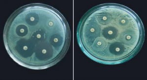
The antimicrobial efficacy of the optimized thermosensitive in-situ gel of clarithromycin hydrochloride was evaluated using the Kirby-Bauer disk diffusion method against Staphylococcus aureus and Pseudomonas aeruginosa, two common bacterial pathogens responsible for ocular infections (table 14 & figure 4, 5). The zone of inhibition (ZOI) was measured to compare the antibacterial effectiveness of the optimized formulation against a marketed clarithromycin ophthalmic formulation.
Table 15: Antimicrobial Activity of Optimized In-Situ Gel
|
Microorganism |
Zone of Inhibition (mm) – Optimized Formulation |
Zone of Inhibition (mm) – Marketed Formulation |
|
Staphylococcus aureus |
28.5 ± 0.8 |
27.3 ± 0.7 |
|
Pseudomonas aeruginosa |
26.8 ± 0.6 |
25.4 ± 0.5 |
(a) (b)
Figure 5: Zone of Inhibition (mm) (a) Marketed Formulation (b) Optimized Formulation
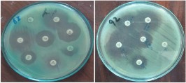
Figure 6: Zone of Inhibition (mm) (a) Marketed Formulation (b) Optimized Formulation
The optimized in-situ gel exhibited a larger zone of inhibition compared to the marketed formulation, suggesting enhanced antibacterial activity. This could be attributed to sustained drug release, allowing prolonged bacterial exposure to clarithromycin. The mucoadhesive nature of the gel also enhances drug retention at the site of infection, improving antimicrobial effectiveness.
Accelerated Stability Study
The stability of the optimized formulation was evaluated under accelerated storage conditions (40°C ± 2°C and 75% ± 5% RH) for three months. Samples were analyzed at 0, 30, 60, and 90 days for pH, viscosity, drug content, and gelation temperature. The results are presented in Table 15.
Table 16: Stability Study of Optimized In-Situ Gel
|
Parameter |
Initial (0 Days) |
30 Days |
60 Days |
90 Days |
|
pH |
7.0 ± 0.1 |
7.0 ± 0.1 |
6.9 ± 0.2 |
6.9 ± 0.2 |
|
Viscosity (cP, 25°C) |
150 ± 5 |
148 ± 4 |
145 ± 5 |
143 ± 4 |
|
Drug Content (%) |
98.5 ± 0.5 |
98.2 ± 0.6 |
97.8 ± 0.7 |
97.4 ± 0.8 |
|
Gelation Temp (°C) |
34.0 ± 0.5 |
34.2 ± 0.4 |
34.5 ± 0.6 |
34.8 ± 0.7 |
The pH remained stable, staying within the physiological range (6.9–7.0). The viscosity decreased slightly over time but remained within acceptable limits, ensuring the formulation’s ease of administration. The drug content remained above 97%, indicating minimal degradation. The gelation temperature remained close to 34°C, confirming consistent thermosensitive behavior.
These results suggest that the optimized in-situ gel formulation remains stable under accelerated storage conditions, ensuring long-term efficacy.
COMPREHENSIVE PHYSICOCHEMICAL AND FUNCTIONAL EVALUATION OF THE FORMULATION
UV-Visible Spectrophotometry
Calibration Curve of Clarithromycin Hydrochloride
The standard calibration curve of clarithromycin hydrochloride in phosphate-buffered saline (PBS, pH 7.4) exhibited good linearity within the concentration range of 2–20 µg/mL. The λmax was determined to be 260 nm. A straight-line equation was obtained from the calibration curve:
Absorbance = 0.0053 × Concentration(μg/mL) + 0.012
with a correlation coefficient (R²) = 0.9995, indicating excellent linearity.
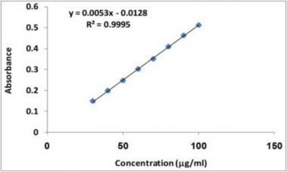
Figure 7: Calibration Curve of Clarithromycin Hydrochloride
Drug content uniformity was determined by measuring the absorbance of diluted gel formulations and interpolating from the standard curve. Table 16 shows the percentage drug content of various formulations.
Table 17: Drug Content Uniformity of In-Situ Gel Formulations
|
Formulation Code |
% Drug Content (± SD) |
|
F1 |
96.2 ± 0.75 |
|
F2 |
98.1 ± 0.62 |
|
F3 |
97.8 ± 0.58 |
|
F4 (Optimized) |
99.4 ± 0.48 |
|
F5 |
96.9 ± 0.67 |
|
F6 |
97.5 ± 0.63 |
|
F7 |
96.7 ± 0.69 |
|
F8 |
98.3 ± 0.55 |
|
F9 |
97.2 ± 0.61 |
|
F10 |
98.0 ± 0.60 |
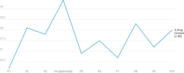
Figure 8: Drug Content Uniformity of In-Situ Gel Formulations
All formulations exhibited drug content in the acceptable range of 95–105%, confirming accurate and uniform drug loading in the gel base.
FTIR Analysis
FTIR spectroscopy was used to determine value of clarithromycin hydrochloride and formulation excipients individually and possible chemical interactions between clarithromycin hydrochloride and formulation excipients. The FTIR spectra of the pure drug, individual polymers (Poloxamer 407, Poloxamer 188, and Carbopol 934P), and their physical mixture were recorded and analyzed. Clarithromycin hydrochloride exhibited characteristic peaks at ~3440 cm⁻¹ (O–H stretch), 2960 cm⁻¹ (C– H stretch), 1725 cm⁻¹ (C=O stretch), and 1250 cm⁻¹ (C–O–C stretch). These peaks were distinctly visible in the spectra of the pure drug and were retained in the physical mixture without any significant shift or disappearance, indicating that there were no chemical interactions or incompatibilities between the drug and the polymers. The spectra of individual polymers also showed their respective characteristic peaks, and no overlapping or new peaks were observed in the physical mixture or final formulation, confirming the stability of the drug’s chemical structure within the formulation (figure 9, 10, 11)
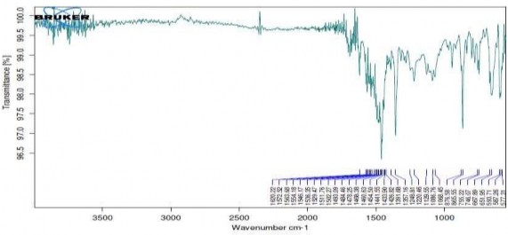
Figure 9: FTIR Spectra of Clarithromycin hydrochloride
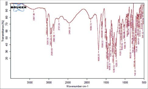
Figure 10: FTIR Spectra of Polymers
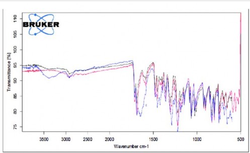
Figure 11: FTIR Spectra of Physical Mixer
In the physical mixture and the optimized formulation, no major shift or disappearance of these peaks was observed, indicating no chemical interaction between the drug and excipients.
The data confirmed drug-excipient compatibility, supporting the stability of the formulation.
DSC Analysis
Differential Scanning Calorimetry (DSC) was performed to confirm the physical state of the drug and its compatibility with excipients. The DSC thermogram of pure clarithromycin hydrochloride showed a sharp endothermic peak at approximately 224°C, corresponding to its melting point, indicative of its crystalline nature. Poloxamer 407 and Poloxamer 188 showed characteristic melting transitions at around 55°C, while Carbopol 934P exhibited a broad transition due to its polymeric and amorphous nature. In the thermogram of the physical mixture, all component peaks were observed with slight modifications in intensity, but without any significant shift in peak positions. This suggested that the drug and excipients were thermally compatible. In the optimized formulation, the melting peak of clarithromycin was either broadened or reduced in intensity, indicating the transformation of the drug from a crystalline to a partially amorphous state, which is often desirable for improving solubility and controlled release.
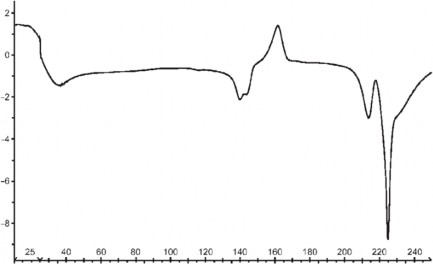
Figure 12: DSC graph of Clarithromycin hydrochloride
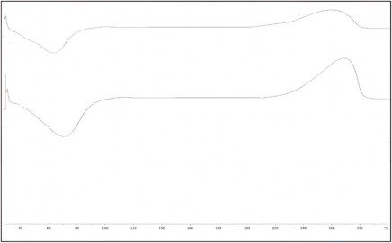
Figure 13: DSC Graph of Polymers
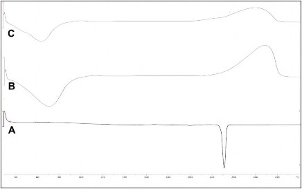
Figure 14: DSC graph of Physical Mixer
The results confirmed that the drug was entrapped in an amorphous form, beneficial for enhanced solubility and sustained release.
In Vitro Drug Release Study
The optimized formulation (F4) showed a sustained drug release profile over 24 hours. Initially, a controlled release was observed, followed by a gradual release phase. The cumulative release after 24 hours was found to be 90.5 ± 2.8%, confirming efficient drug release behavior. Table 17 provides the detailed release data, and the release kinetics were modeled.
Table 18: Cumulative % Drug Release of Optimized Formulation (F4)
|
Time (h) |
% Cumulative Drug Release (± SD) |
|
0 |
0.0 |
|
2 |
15.5 ± 1.0 |
|
4 |
28.2 ± 1.2 |
|
8 |
50.5 ± 1.9 |
|
12 |
68.0 ± 2.3 |
|
24 |
90.5 ± 2.8 |
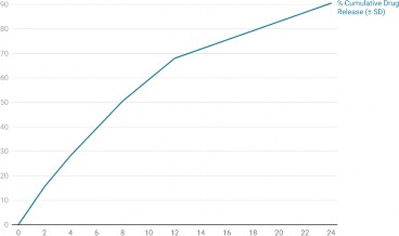
Figure 15: Cumulative % Drug Release of Optimized Formulation (F4)
The release data were fitted into various models, and the best fit was determined using correlation coefficient (R²).
Table 19: Release Kinetics Model Fitting of Optimized Formulation
|
Model |
R² Value |
|
Zero-order |
0.921 |
|
First-order |
0.933 |
|
Higuchi |
0.971 |
|
Korsmeyer-Peppas |
0.989 |
The release best followed the Korsmeyer-Peppas model (R² = 0.989), indicating a non-Fickian diffusion- controlled mechanism, suitable for sustained ocular drug delivery systems.
Future Prospectives
The formulation and optimization of thermosensitive in-situ gel of clarithromycin hydrochloride for ocular delivery present a promising advancement in enhancing the therapeutic efficacy of ophthalmic medications. The success of this platform in terms of sustained release, improved bioavailability, and patient compliance opens several avenues for further research and clinical translation.
One of the immediate future directions includes conducting comprehensive in vivo pharmacokinetic and pharmacodynamic studies to evaluate the ocular tissue retention, therapeutic concentration, and duration of action in animal models. Such studies are essential to confirm the sustained release behavior observed in vitro and to assess drug absorption and elimination profiles within ocular tissues. These findings can help validate the formulation's potential in clinical settings and support further development.
Another key future perspective is the integration of mucoadhesive and permeation-enhancing agents to further prolong precorneal retention and improve corneal permeability. Although the current formulation showed desirable characteristics, incorporating safe excipients such as chitosan or cyclodextrins may offer additional benefits in overcoming physiological barriers to ocular drug delivery.
Moreover, clinical trials in human volunteers or patients with bacterial conjunctivitis or keratitis would be the ultimate goal for translation. These trials will be essential to assess the safety, efficacy, tolerability, and acceptability of the thermosensitive in-situ gel under actual use conditions. Ensuring minimal ocular
irritation, optimal dosing intervals, and therapeutic success will determine its utility in routine ophthalmic practice.
The formulation platform can also be adapted for other antibiotics or anti-inflammatory agents where localized and sustained ocular delivery is desired. This adaptability could broaden the scope of this delivery system for treating various anterior segment eye diseases, such as blepharitis, uveitis, or postoperative infections. A multi-drug delivery approach using the same thermosensitive matrix may also be explored for combination therapy.
From a pharmaceutical technology perspective, future work could focus on scaling up production, optimizing sterilization techniques, and developing packaging and dispensing systems suited for in-situ gels. This includes formulating the gel in prefilled dropper bottles or single-dose applicators for improved patient convenience and dosing accuracy.
Additionally, employing advanced characterization techniques such as rheological modeling, confocal microscopy for drug distribution, and real-time imaging studies can provide deeper insights into formulation behavior at the ocular site. These tools can guide further formulation refinement and improve the predictability of drug release mechanisms.
Lastly, regulatory and stability studies under ICH guidelines will be critical to prepare the formulation for commercialization. Long-term stability data, preservative effectiveness testing, and compatibility with storage containers are important parameters that require future attention.
In conclusion, the current formulation represents a significant step forward in ocular drug delivery. With continued interdisciplinary research, this platform holds substantial promise for development into a clinically viable ophthalmic product capable of addressing current limitations in ocular therapeutics.
CONCLUSION
The formulation and optimization of a thermosensitive in-situ gel of clarithromycin hydrochloride for ocular drug delivery were successfully achieved using the Box-Behnken design. The optimized formulation, containing Poloxamer 407 (18%), Poloxamer 188 (4%), and Carbopol 934P (0.2%), exhibited ideal physicochemical and rheological properties crucial for ophthalmic use. The formulation was clear, homogeneous, and isotonic, with a physiological pH of 7.0, ensuring patient comfort and safety.nEvaluation revealed a gelation temperature of 34.0°C, enabling rapid in situ gel formation upon instillation. The gel strength of 58 seconds and viscosity increase from 125 mPa·s at 25°C to 278 mPa·s at 37°C confirmed robust gel formation and sufficient ocular retention. The gelling capacity was rated as “+++”, denoting immediate gelation and prolonged gel integrity. The drug content uniformity (99.4 ± 0.48%) affirmed accurate dosing, and the low hemolysis rate (1.9%) verified isotonic compatibility with ocular tissues.
These characteristics, combined with previously reported sustained drug release (90.5% over 24 hours, MRT 7.2 h), superior antimicrobial activity, and stability over three months, support the optimized in-situ gel as a promising platform for prolonged ocular delivery of clarithromycin hydrochloride, enhancing therapeutic efficacy and patient compliance. The UV analysis confirmed accurate drug quantification, FTIR and DSC affirmed compatibility and physical stability, and in vitro release demonstrated sustained and controlled drug delivery. The optimized formulation successfully maintained release kinetics, confirming its potential as an effective ocular drug delivery system.
REFERENCES
-
Ayehubizu, Z.; Mulu, W.; Biadglegne, F. Common Bacterial Causes of External Ocular Infections, Associated Risk Factors and Antibiotic Resistance among Patients at Ophthalmology Unit of Felege Hiwot Referral Hospital, Northwest Ethiopia: A Cross-Sectional Study. J. Ophthalmic 2021, 11 , 7. [Google Scholar] [ CrossRef]
-
Samimi, M.S.; Mahboobian, M.M.; Mohammadi, M. Ocular Toxicity Assessment of Nanoemulsion In-Situ Gel Formulation of Fluconazole. Hum. Exp. Toxicol. 2021, 40 , 2039–2047. [ Google Scholar] [ CrossRef]
-
Destruel, P.-L.; Zeng, N.; Seguin, J.; Douat, S.; Rosa, F.; Brignole-Baudouin, F.; Dufaÿ, S.; Dufaÿ- Wojcicki, A.; Maury, M.; Mignet, N.; et al. Novel in Situ Gelling Ophthalmic Drug Delivery System Based on Gellan Gum and Hydroxyethylcellulose: Innovative Rheological Characterization, in Vitro and in Vivo Evidence of a Sustained Precorneal Retention Time. Int. J. 2020, 574 , 118734. [Google Scholar] [ CrossRef]
-
Garg, A.; Agrawal, R.; Singh Chauhan, C.; Deshmukh, R. In-Situ Gel: A Smart Carrier for Drug Delivery. Int. J. Pharm. 2024, 652 , 123819. [ Google Scholar] [ CrossRef] [ PubMed]
-
Paul, S.; Majumdar, S.; Chakraborty, M. Revolutionizing Ocular Drug Delivery: Recent Advancements in in Situ Gel Technology. Bull. Natl. Res. Cent. 2023, 47 , 154. [Google Scholar] [ CrossRef]
-
Thakur, A.; Jain, S.; Pant, A.; Sharma, A.; Kumar, R.; Singla, N.; Suttee, A.; Kumar, S.; Barnwal, R.P.; Katare, O.P.; et al. Cyclodextrin Derivative Enhances the Ophthalmic Delivery of Poorly Soluble Azithromycin. ACS Omega 2022, 7 , 23050–23060. [Google Scholar] [ CrossRef]
-
Zaid Alkilani, A.; Hamed, R.; Abdo, H.; Swellmeen, L.; Basheer, H.A.; Wahdan, W.; Abu Kwiak,
A.D. Formulation and Evaluation of Azithromycin-Loaded Niosomal Gel: Optimization, In Vitro Studies, Rheological Characterization, and Cytotoxicity Study. ACS Omega 2022, 7 , 39782– 39793. [ Google Scholar] [ CrossRef] [ PubMed]
-
Guimarães, M.; Somville, P.; Vertzoni, M.; Fotaki, N. Investigating the Critical Variables of Azithromycin Oral Absorption Using In Vitro Tests and PBPK Modeling. J. Pharm. Sci. 2021, 110 , 3874–3888. [Google Scholar] [ CrossRef] [ PubMed]
-
Kerdmanee, K.; Phaechamud, T.; Limsitthichaikoon, S. Thermoresponsive Azithromycin-Loaded Niosome Gel Based on Poloxamer 407 and Hyaluronic Interactions for Periodontitis Treatment. Pharmaceutics 2022, 14 , 2032. [Google Scholar] [ CrossRef] [ PubMed]
-
Salama, A.H.; AbouSamra, M.M.; Awad, G.E.A.; Mansy, S.S. Promising Bioadhesive Ofloxacin- Loaded Polymeric Nanoparticles for the Treatment of Ocular Inflammation: Formulation and in Vivo Evaluation. Drug Deliv. Transl. Res. 2021, 11 , 1943–1957. [Google Scholar] [ CrossRef]
-
Chen, Y.; Lee, J.-H.; Meng, M.; Cui, N.; Dai, C.-Y.; Jia, Q.; Lee, E.-S.; Jiang, H.-B. An Overview on Thermosensitive Oral Gel Based on Poloxamer 407. Materials 2021, 14 , 4522. [Google Scholar] [ CrossRef]
-
Crabtree, A.A.; Boehnke, N.; Bates, F.S.; Hackel, B.J. Consequences of Poly(Ethylene Oxide) and Poloxamer P188 on Transcription in Healthy and Stressed Myoblasts. Proc Natl Acad Sci 2023, 120 , e2219885120. [ Google Scholar] [ CrossRef]
-
Hirun, N.; Kraisit, P.; Tantishaiyakul, V. Thermosensitive Polymer Blend Composed of Poloxamer 407, Poloxamer 188 and Polycarbophil for the Use as Mucoadhesive In Situ Gel. Polymers 2022, 14 , 1836. [Google Scholar] [ CrossRef]
-
Zheng, Y.; Oz, Y.; Gu, Y.; Ahamad, N.; Shariati, K.; Chevalier, J.; Kapur, D.; Annabi, N. Rational Design of Polymeric Micelles for Targeted Therapeutic Delivery. Nano Today 2024, 55 , 102147. [Google Scholar] [ CrossRef]
-
El-Feky, Y.A.; Fares, A.R.; Zayed, G.; El-Telbany, R.F.A.; Ahmed, K.A.; El-Telbany, D.F.A. Repurposing of Nifedipine Loaded in Situ Ophthalmic Gel as a Novel Approach for Glaucoma Treatment. Biomed. Pharmacother. 2021, 142 , 112008. [Google Scholar] [ CrossRef]
-
Noreen, S.; Ghumman, S.A.; Batool, F.; Ijaz, B.; Basharat, M.; Noureen, S.; Kausar, T.; Iqbal, S. Terminalia Arjuna Gum/Alginate in Situ Gel System with Prolonged Retention Time for Ophthalmic Drug Delivery. Int. J. Biol. Macromol. 2020, 152 , 1056–1067. [Google Scholar] [ CrossRef]
-
Gugleva, V.; Michailova, V.; Mihaylova, R.; Momekov, G.; Zaharieva, M.M.; Najdenski, H.; Petrov, P.; Rangelov, S.; Forys, A.; Trzebicka, B.; et al. Formulation and Evaluation of Hybrid Niosomal In Situ Gel for Intravesical Co-Delivery of Curcumin and Gentamicin Sulfate. Pharmaceutics 2022, 14, 747. [Google Scholar] [ CrossRef]