
GASTRODISCOIDIASIS: SYSTEMATIC-NARRATIVE HYBRID LITERATURE REVIEW
Paulo Sérgio Balbino Miguel1*; Ademir Nunes Ribeiro Júnior2; Lucas Brandão-Gonçalves3; Tiago Ricardo Moreira4; Andréia Patrícia Gomes5
1 Federal Institute of Paraná, Campus Barracão, Paraná, Brazil; Orcid ID: https://orcid.org/0000- 0002-6652-366X; 2 Evangelical University of Goiás, Anápolis, Brazil; Orcid ID: https://orcid.org/0000-0003-1661-347X; 3 Federal Institute of Paraná, Campus Barracão, Paraná, Brazil; Orcid ID: https://orcid.org/0009-0007-2650-6674; 4 Federal University of Viçosa, Viçosa, Brazil; Orcid ID: https://orcid.org/0000-0002-6606-4942; 5 Federal University of Rio de Janeiro, Rio de Janeiro, Brazil; Orcid Id: https://orcid.org/0000-0002-5046-6883;
Corresponding Author: Paulo Sérgio Balbino Miguel Email: paulo.miguel@ifpr.edu.br
ABSTRACT
Gastrodiscoidiasis is among the neglected, underreported, and difficult-to-diagnose intestinal diseases. Human infection occurs through the ingestion of raw or undercooked fish or vegetables contaminated with the metacercaria of the helminth. In this study, we analyzed the clinical and epidemiological profile of Gastrodiscoidiasis and identified the main diagnostic methods used in documented cases of the disease. The reviewed cases revealed that the recognition of the infection is hindered by nonspecific clinical manifestations, especially in cases of coinfection with other helminthiases. Most reports of the disease come from India; however, the pathogen has also been described in the United States, Canada, Indonesia, Ghana, and the Philippines, though without reports of clinical disease. The main drugs used in treatment are praziquantel and mebendazole. Gastrodiscoidiasis should be considered as a differential diagnosis in patients with gastrointestinal symptoms and a history of dietary exposure in endemic areas, including travelers. Greater efforts are needed to strengthen the clinical and epidemiological investigation of this disease.
Keywords: Intestinal trematode, Gastrodiscoidiasis, Gastrodiscoides hominis
INTRODUCTION
Gastrodiscoidiasis is a foodborne zoonotic disease caused by the trematode Gastrodiscoides [1]. Although infections are often asymptomatic, the presence of the parasite may lead to clinical manifestations such as diarrhea, malnutrition, and, in severe cases, death. The
pathogen is most prevalent in India [2][3], with additional reports in Africa [4] and Russia [5]. Humans become infected after ingesting raw or undercooked fish or vegetables contaminated with the parasite’s metacercaria. In the small intestine of the definitive host, the metacercaria develop into hermaphroditic adult worms, which then migrate to the cecum and ascending colon, where they deposit hundreds of eggs [6]. The eggs, once excreted in feces and reaching bodies of water, hatch within 16 to 17 days at temperatures between 26°C and 32°C [7], releasing the free-swimming larval form known as a miracidium, which can infect the snail Helicorbis coenosus . Within this intermediate host, the parasite progresses through sporocyst, redia, and cercaria stages. Once released into aquatic environments, cercariae can contaminate plants, crustaceans, amphibians, mollusks, and fish [6] (Figure 1). The incubation period and symptomatology of Gastrodiscoidiasis remain poorly understood. However, repeated ingestion of cercariae or metacercaria results in more severe infections and, consequently, more evident clinical complaints [3][6].
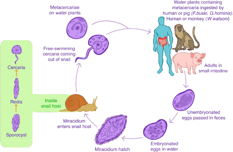
Figure 1. Biological cycle of Gastrodiscoides hominis during human
Gastrodiscoidiasis.
Image created by Ademir Nunes Ribeiro Júnior.
One of the first detailed endoscopic reports described a 15-year-old adolescent with chronic abdominal pain, in whom colonoscopy revealed multiple 8–10 mm worms adhered to the cecal mucosa; removal of the parasites caused local bleeding. The severity of the disease is generally associated with the number of parasites attached to the intestinal tract, leading to mucosal inflammation [2]. In humans, Gastrodiscoidiasis predominantly occurs in rural areas of endemic countries, where poor sanitation, the use of contaminated water, and the consumption of aquatic vegetables or raw or undercooked mollusks create favorable conditions not only for the life cycle of G. hominis but also for other foodborne helminths [8][9]. In addition to local factors, global phenomena such as globalization and climate change may significantly influence the dynamics and epidemiological patterns of these infections. Increased human mobility and international food trade, combined with temperature and precipitation shifts, facilitate the introduction of intermediate hosts into new regions and may sustain transmission cycles in previously non-endemic areas [8].
Furthermore, changes in dietary habits, the expansion of global markets, improved transportation systems, and population growth increase the number of individuals at risk of infection [10]. This context underscores the need for a better understanding of Gastrodiscoidiasis, considered one of the most neglected foodborne intestinal trematodiases worldwide [6][11]. This study aims to analyze the clinical and epidemiological profile of
human Gastrodiscoidiasis and to identify and describe the main diagnostic methods used in documented cases of the disease.
METHOD
This study employed the hybrid review approach proposed by [12], combining a narrative methodology aligned with the research objectives and a systematic strategy to support an exhaustive literature search, using detailed inclusion and exclusion criteria for article selection. This narrative-systematic strategy has been widely adopted in health research [13][14][15], allowing for more focused and comprehensive searches, with pre-established critical criteria and quantitative description of the synthesized data [12]. This choice also enabled the presentation of results along with a discussion of the methods used in the literature search for this review, which is guided by the following research question: What are the clinical and epidemiological profiles of human Gastrodiscoidiasis, and what are the main diagnostic methods used?
Eligibility Criteria:
Eligible studies included original research with primary data, involving human populations infected with Gastrodiscoidiasis. Studies were not excluded if the infection was acquired through the ingestion of contaminated vegetables or other food sources, or if they reported mixed infections in addition to G. hominis . Both incident and prevalent cases were considered, including those with definitive diagnoses (confirmed through traditional parasitological methods such as Kato-Katz or standard stool examination), presumptive diagnoses (based on clinical or imaging criteria), or diagnoses involving molecular biology techniques.
Studies were excluded if they focused on animal populations other than humans, addressed topics unrelated to this research, or were based solely on secondary data. No restrictions were applied regarding language, year of publication, or geographic location. Articles published in languages other than English or Portuguese were translated using the “DeepL” tool and the artificial intelligence tool “Sider Fusion.” Only studies published up to April 2025 were included.
Information Sources and Search Strategy:
The databases selected for article retrieval were: Web of Science, PubMed, Scopus, ScienceDirect, Primo bibTex, Regional Portal of the Virtual Health Library, EMBASE, and EBSCO. Additionally, searches were conducted in Google Scholar and the gray literature. The search strategy, conducted in April 2025, used the following MeSH terms: “( Gastrodiscioides ) OR (Gastrodiscoidiasis) OR (Gastrodiscoidiasis) AND (Humans) OR (Man) OR (Human) OR ( Homo sapiens ).” The reference lists of relevant articles were also reviewed to identify additional potentially eligible studies. The Rayyan software was used to manage the references.
In Rayyan, all electronically identified articles were uploaded, organized, and screened to remove duplicates. Titles and abstracts were reviewed based on the eligibility criteria. When titles and abstracts lacked sufficient data for assessment, full-text articles were retrieved and reviewed. Only studies involving humans infected with G. hominis were included.
A data extraction protocol was developed for the selected full-text articles. Extracted and categorized data included: author/year, mean patient age, geographic region of origin, number of positive cases, signs and symptoms of the disease, diagnostic methods, and treatment.
RESULTS
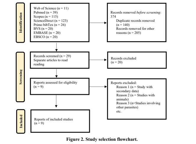
A total of 374 articles were identified from the following databases: Web of Science, PubMed, Scopus, ScienceDirect, Primo bibTex, Regional Portal of the Virtual Health Library, EMBASE, and EBSCO. Of these, 140 were excluded due to duplication, and another 205 were excluded after reading titles and abstracts. These exclusions were based on the use of secondary data, unrelated topics (involving other helminths), or studies focusing on non-human animals. During the initial assessments, 29 full-text articles were reviewed, of which nine were included in this study for meeting the established inclusion criteria (Figure 2). The selected studies investigated 152 individuals diagnosed with Gastrodiscoidiasis—150 of them in India, one in Nigeria, and one in Nepal. Detailed information on the selected articles for this hybrid review is provided in Table 1. All included studies were case reports.
Table 1. Identification of selected articles for the hybrid literature review on human Gastrodiscoidiasis.
|
Author / Year |
Mean Age |
Region of Origin |
Number of Positive Cases |
Signs & Symptoms |
Diagnosis |
Treatment |
|
[2] |
15 |
India |
1 |
Periumbilical and discomforting abdominal pain; contracted cecum with irregular contours |
Colonoscopy, barium study, parasite histology |
Mebendazole 100 mg, twice daily for 3 days; treatment repeated after colonoscopy with hypertonic saline flushing of parasites; patient cured |
|
[3] * |
20 |
India |
1 |
Pedal edema lasting two months; nausea; abdominal discomfort; hyperpigmented brownish rashes on both legs |
Endoscopy; colonoscopy; stool examination (SPE); blood test; ultrasound; Doppler; abdominal ultrasonograp hy. Findings: anemia, low total protein, mild ascites, diffuse soft tissue edema; infestation in the terminal ileum and right colon |
Praziquantel (25 mg/kg, single dose), albendazole (400 mg/day for 3 days), and metronidazole (400 mg every 8 hours for 5 days) |
|
[16] |
Young adult |
India |
1 |
Excess mucus in stool for three months |
Negative stool parasitologic al exam (SPE); colonoscopy performed; patient did not return for repeat colonoscopy |
Praziquantel (no clinical response) |
|
[4] |
66 |
Nepal |
1 |
Upper abdominal pain for 4 months; history of black stools; anemia; eosinophilia; bloody stools |
Upper gastrointestin al endoscopy; stool parasitologic al exam (SPE); peripheral blood smear; findings: anemia and eosinophilia |
Praziquantel, three doses (25 mg/kg each) in one day, followed by purgatives; patient improved |
|
[7] * |
Children (87%) |
India |
91 |
- |
Stool parasitologic al exam (SPE) |
Beta-naphthol; soap water enemas (clysters) |
|
[17] |
7,5 |
Nigeria |
1 |
Malnutrition; anemia; occasional diarrhea with mucus; increased splenic circumference; moderately pale; diffuse generalized lymphadenopathy ; mild hepatosplenomeg aly; marasmus; eosinophilia; low white blood cell count |
Kato-Katz; a stool exam repeated weekly over three weeks showed complete elimination of both parasites |
Single oral dose of 500 mg mebendazole |
|
[18] * |
18 a 40 / 10 a 50 |
India |
56 |
Weakness; loss of appetite; colitis; dysentery |
Stool parasitologic al exam (SPE) |
Thymol |
* Case reports involving mixed infections
** The case studies by [19] & [20] are not included in this table, as they address vegetables contaminated with G. hominis eggs
The symptoms of Gastrodiscoidiasis are varied and nonspecific. Among the reported manifestations are: periumbilical abdominal pain [2], pedal edema, nausea, abdominal discomfort, hyperpigmented brownish skin rashes on the legs [3], excess mucus in the stool [16], upper abdominal pain with a history of dark-colored stools, anemia, and eosinophilia [4], malnutrition, occasional diarrhea with mucus, increased splenic circumference, diffuse generalized lymphadenopathy, mild hepatosplenomegaly, and low white blood cell count [17], weakness, loss of appetite, colitis, and dysentery [18]. It is important to note that some of these symptoms were recorded in mixed infections with other parasitic diseases (Table 1), such as those described by [3] (amoebiasis, giardiasis, and fasciolopsiasis), [7] (ascariasis, trichuriasis, fasciolopsiasis, and ancylostomiasis), and [18] (ascariasis, trichuriasis, ancylostomiasis, enterobiasis, fasciolopsiasis, and strongyloidiasis).
Symptoms such as pedal edema, nausea, abdominal discomfort, hyperpigmented brownish leg rashes [3], and weakness, loss of appetite, colitis, and dysentery [18] were likely influenced by the presence of other pathogens. The most frequently reported symptom across the reviewed studies was abdominal pain, as identified by [2,3,4]. The presence of this and other nonspecific clinical signs highlights the importance of laboratory testing for more accurate diagnosis of the disease.
Colonoscopy proved to be the most effective method in demonstrating the presence of the pathogen [2,3,16]. Indeed, G. hominis typically resides in the cecum and ascending colon of both humans and animals [21], which may explain the high diagnostic efficiency of colonoscopy for this infection. Conversely, stool parasitological examination (SPE), the most commonly used diagnostic method among the reviewed studies, yielded negative results in most cases. However, when positive, SPE enabled the identification of up to 13,500 eggs [17]. In adult patients, the use of soap water enemas allowed for the recovery of 989 worms, while treatment with beta-naphthol proved ineffective [7].
One effective treatment regimen involved mebendazole 100 mg, administered twice daily for three days, followed by a second colonoscopy and topical application of hypertonic saline solution to the parasites [2]. Another successful treatment protocol consisted of praziquantel in three doses (25 mg/kg each) administered in a single day, followed by the use of purgatives [4]. The use of other drug regimens may have been hindered by patient nonadherence to treatment.
DISCUSSION
In this hybrid review, we explored the clinical and epidemiological profile of Gastrodiscoidiasis and the main diagnostic methods used for this neglected and understudied disease in humans, which is transmitted through the ingestion of metacercariae present in aquatic plants, fish, crayfish, squid, and undercooked mollusks [22]. The signs and symptoms of Gastrodiscoidiasis are nonspecific, which hinders early clinical diagnosis. In humans, the pathogen causes inflammatory reactions in the intestinal mucosa, particularly in the ascending colon [22], ileum [2], and cecum, resulting in diarrhea [22]. In some cases, the cecal region becomes so heavily infected that visualization of healthy tissue is difficult [23]; such tissue may exhibit superficial desquamation, eosinophilic and lymphocytic infiltration [24], and diffuse soft tissue edema [3]. It is important to note, however, that in the latter case, the patient was also diagnosed with other parasitic infections (e.g., Entamoeba histolytica , Giardia , and Fasciola hepatica ). Therefore, the health complications arising from G. hominis infection must be investigated, especially given the documented deaths among untreated patients, particularly children [22].
Most (59.8%) of the patients described in the eligible studies had consumed aquatic plants. These patients, mainly Indian children, were treated with soap water enemas, which released pale brownish-green rhomboid-shaped eggs, measuring 150–170 μm in length and 60–70 μm in width [7]. The eggs of G. hominis are operculated, oval, and possess a thickened abopercular end, with an average size of 146 μm long and 66 μm wide [25]. Detection of these eggs in the feces of infected individuals is generally considered the standard laboratory criterion for
diagnosing Gastrodiscoidiasis [6]. However, in this review, most stool parasitological examinations were negative. When positive, the Kato-Katz technique allowed the detection of thousands of eggs in a single individual [17]. Could Kato-Katz be the most effective method for diagnosing Gastrodiscoidiasis? It is also essential to follow recommended guidelines for stool sample collection—examining three or more samples collected on different days and preserved or refrigerated until analysis [26]—as failure to do so may account for false negatives. Moreover, negative results may be due to testing performed outside the egg- shedding period of mature worms. The effectiveness of colonoscopy in demonstrating the presence of the pathogen must also be highlighted. In fact, G. hominis resides in the cecum and ascending colon in both humans and animals [21], which may explain the diagnostic value of colonoscopy for this infection.
Praziquantel appears to be the preferred medication [22], and it was prescribed in the eligible studies included in this hybrid review. However, evaluating its efficacy proved difficult, as patients did not return for follow-up examinations [16]. One effective treatment involved mebendazole 100 mg, administered twice daily for three days, repeated after a second colonoscopy, followed by flushing of the parasites with hypertonic saline solution [2]. Another successful protocol used praziquantel in three doses (25 mg/kg each) in a single day, followed by purgatives [4]. Nonetheless, these case studies involved only one patient each, and more research is needed to establish a specific treatment regimen for G. hominis infection.
Among endemic regions, India has the highest prevalence, particularly in remote rural and semi-urban areas. Other countries with reported cases include Pakistan, the Philippines, Vietnam [27], Nepal [4], and Nigeria [17]. However, in this review, no primary studies from the first three countries were found (Table 1). In all these countries, the data are likely underestimated, as Gastrodiscoidiasis is often asymptomatic [6]. Additionally, reports of the pathogen’s presence in the United States, Canada, Indonesia, Ghana, and the Philippines [28], and evidence of similar transmission pathways in animals [27], suggest a broader but underreported distribution. Alongside underreporting, a major methodological gap remains: there are no spatial distribution studies for G. hominis . Schluth et al. [11] showed that out of 232 helminth species known to infect humans, only 45 (<20%) have any form of cartographic representation, mostly focusing on schistosomiasis, filariasis, hookworm, ascariasis, and trichuriasis. Foodborne trematodes such as G. hominis remain unmapped. This lack of geospatial data hinders the identification of priority areas for surveillance and control, as well as accurate estimates of prevalence and disease burden.
Furthermore, G. hominis eggs have been found on cabbage in Nigeria [19,20], reinforcing the possibility of foodborne transmission, as it is through this parasitic form that metacercariae encyst in food [22]. Therefore, Gastrodiscoidiasis should be considered a possible diagnosis in patients with intestinal symptoms, particularly during health screening of travelers returning from endemic areas and in parasitological surveys. In endemic regions, patients should also be educated about prophylactic measures such as proper washing of aquatic plants and consumption of treated water [21].
CONCLUSIONS
Gastrodiscoidiasis remains a neglected intestinal helminthiasis, difficult to diagnose and frequently underreported. The cases analyzed in this review reveal nonspecific clinical manifestations such as abdominal pain, diarrhea, anemia, and eosinophilia, which hinder the recognition of the infection, particularly in settings of coinfection. Although the presence of eggs in feces is considered the primary diagnostic criterion, it has shown limited sensitivity, possibly due to inadequate sample collection, improper techniques, or low parasitic burden. Despite the frequent use of praziquantel and mebendazole, data on their therapeutic efficacy
are still limited and based on isolated case reports. Therefore, there is a need to strengthen clinical and epidemiological investigations of this disease, with an emphasis on more sensitive diagnostic protocols and clinical studies that validate effective treatment regimens. Gastrodiscoidiasis should be considered a differential diagnosis in patients with gastrointestinal symptoms and a history of dietary exposure in endemic areas.
REFERENCES
-
Chai, J. Y., & Jung, B. K. (2020). Foodborne intestinal flukes: A brief review of epidemiology and geographical distribution. Acta Tropica , 201 , 105210.
-
Gupte, A., Shah, C., Koticha, A., Shukla, A., Kuyare, S., & Bhatia, S. (2014). Gastrodiscoides hominis infestation of colon: endoscopic appearance. Gastrointestinal , 79 (4), 549-550.
-
Sunil, H. S., Gandhi, B. P., Avinash, B., Devi, G., & Sudhir, U. (2014). Parasitic Zoo of Fasciolopsis buski, Gastrodiscoides hominis , Giardia intestinalis and Entamoeba histolytica. Intern. Med , 4 , 135.
-
Sah, R., Acosta, L., & Toledo, R. (2019). A case report of human Gastrodiscoidiasis in Nepal. Parasitology International , 71 , 56-58.
-
Kondrashin, A. V., Morozova, L. F., Stepanova, E. V., Turbabina, N. A., Maksimova, M. S., Anikina, A. S., ... & Morozov, E. N. (2023). A rare human helminth infection in Russia. Tropical Medicine and Infectious Disease , 8 (8), 403.
-
Toledo, R., Álvarez-Izquierdo, M., Esteban, J. G., & Muñoz-Antoli, C. (2022). Neglected food-borne trematodiases: echinostomiasis and Gastrodiscoidiasis. Parasitology , 149 (10), 1319-1326.
-
Buckley, J. J. C. (1939). Observations on Gastrodiscoides hominis and Fasciolopsis buski in Assam. Journal of Helminthology , 17 (1), 1-12.
-
Pozio, E. (2020). How globalization and climate change could affect foodborne parasites. Experimental Parasitology, 208, 107807.
-
Chai, JY., Jung, BK. (2019). Epidemiology of Trematode Infections: An Update. In: Toledo, R., Fried, B. (eds) Digenetic Trematodes. Advances in Experimental Medicine and Biology, vol 1154. Springer, Cham. https://doi.org/10.1007/978-3-030-18616-6_12
-
Chávez-Ruvalcaba F, Chávez-Ruvalcaba MI, Moran Santibañez K, Muñoz-Carrillo JL, León Coria A, Reyna Martínez R. Foodborne Parasitic Diseases in the Neotropics – A Review. Helminthologia. 2021 Jun 8;58(2):119-133. Doi: 10.2478/helm-2021-0022. PMID: 34248373; PMCID: PMC8256457.
-
Schluth CG, Standley CJ, Bansal S, Carlson CJ. Spatial parasitology and the unmapped human helminthiases. Parasitology. 2023;150(4):391-399. doi:10.1017/S0031182023000045
-
Turnbull, D., Chugh, R., & Luck, J. (2023). Systematic-narrative hybrid literature review: A strategy for integrating a concise methodology into a manuscript. Social Sciences & , 7 (1), 100381.
-
Rusli, K. D. B., Chua, W. L., Ang, W. H. D., Ang, S. G. M., Lau, Y., & Liaw, S. Y. (2024).
A hybrid systematic narrative review of instruments measuring home‐based care nurses'
competency. Journal of Advanced Nursing , 80 (7), 2647-2671.
-
De, G., Sondhi, N., Bhattacharjee, A., & Joshi, H. (2024). Preventive healthcare behavior: A hybrid systematic literature review (1998–2023). International Journal of Consumer , 48 (1), e13000.
-
Khan, N., Kurnik-Łucka, M., Latacz, G., & Gil, K. (2024). Systematic-Narrative Hybrid Literature Review: Crosstalk between Gastrointestinal Renin–Angiotensin and Dopaminergic Systems in the Regulation of Intestinal Permeability by Tight Junctions. International Journal , 25 (10), 5566.
-
Shukla, S. K., Asati, P. K., Banerjee, T., & Dixit, V. K. (2016). Rare trematode infestation in irritable bowel syndrome: Pathogen or commensal? Journal of Digestive Endoscopy , 7 (03), 123-124.
-
Dada-Adegbola, H. O., Falade, C. O., Oluwatoba, O. A., & Abiodun, O. O. (2004). Gastrodiscoides hominis infection in a Nigerian-case report. West African Journal of , 23 (2), 185-186.
-
Sharma, A. N. (1930). Helminthic infections in Shillong. The Indian Medical , 65 (4), 200.
-
Uzairue, L. I., Ugbor, C. I., Ezeah, G. A., Eze, N. O., & Nwadike, I. G. (2013). Gastrodiscoides hominis infestation on vegetables (cabbages) sold in Ekpoma Markets, Edo State, Southern Nigeria-a case report. International Journal of Basic, Applied and Innovative , 2 (2), 37-39.
-
Okpala, H. O., Uzairue, L. I., Akhile, A., Aghatise, K., & Digban, K. (2016). Determination of human parasitic infestation on fruits and vegetables sold in Ekpoma markets, Edo State, Nigeria. Asian Journal of Medical Sciences , 8 (3), 10-14.
-
Barriga O. O. Edition, T. (2003). ZOONOSES AND COMMUNICABLE DISEASES
COMMON TO MAN AND ANIMALS. Disponível em chrome- extension://efaidnbmnnnibpcajpcglclefindmkaj/https://cdn.who.int/media/docs/default- source/ntds/echinococcosis/zoonosesvol-3.pdf?sfvrsn=7a04da2a_4&download=true
-
Mas-Coma, S., Bargues, M. D., & Valero, M. A. (2006). Gastrodiscoidiasis, a plant-borne zoonotic disease caused by the intestinal amphistome fluke Gastrodiscoides hominis (Trematoda: Gastrodiscoidiasis). Rev Iber Parasitol , 66 , 75-81.
-
Shrivastav, H. O., & Shah, H. L. (1970). On Gastrodiscoides hominis (Lewis and McConnell, 1876) Leiper, 1913 from pigs 9 (Sus scrofa domestica) in Madhya Pradesh, its pathology and public health importance. Indian Journal of Pathology & Bacteriology , 13 (2), 68-72.
-
Ahluwalia, S. S. (1960). Gastrodiscoides hominis (Lewis and McConnell) Leiper 1913 (the amphistome parasite of man and pig).
-
Dutt, S. C., & Srivastava, H. D. (1972). The life history of Gastrodiscoides hominis (Lewis and McConnel, 1876) Leiper, 1913–the amphistome parasite of man and pig. Journal of , 46 (1), 35-46.
-
National Center for Emerging and Zoonotic Infectious Diseases (NCEZID). Diagnosis of Parasitic Diseases. April 9, 2024. 27. https://www.cdc.gov/parasites/testing- diagnosis/index.html. Acesso em 12/04/2025
-
Buckley, J. J. C. (1964). The Problem of Gastrodiscoides hominis (Lewis and McConnell, 1876) Leiper, 1913. Journal of Helminthology, 38(1-2), 1-6.
-
Orrell T, Informatics and Data Science Center - Digital Stewardship (2025). NMNH Extant
Specimen Records (USNM, US). Version 1.92. National Museum of Natural History, Smithsonian Institution. Occurrence dataset https://doi.org/10.15468/hnhrg3 accessed via GBIF.org on 2025-04-14.